This Article
The Role of Amnion Membrane-Derived Mesenchymal Stem Cells on Differentiation and Expansion of Natural Killer Cell Progenitors Originated From Umbilical Cord Blood Mononuclear Cells
Abstract
Background: Natural killer (NK) cells are members of the innate immune system. Their unique properties, including recognition of viral infected and tumor cells without major histocompatibility complex (MHC) restriction or prior sensitization, make them a suitable choice for immunotherapy. Low numbers of NK cells in circulating blood is the most important obstacle for this goal.
Objectives: The aim of this study was to make an optimum in vitro condition to proliferate and differentiate cord blood (CB)-NK cell progenitors to mature NK cells, which can be used for cell therapy.
Materials and Methods: In our study, CB-Mononuclear Cells’ (MNCs) CD3+ lymphocytes were positive depleted using immunomagnetic microbeads. This CD3-depleted (CD3-dep) CB – MNCs compartment was used for in vitro expansion with or without a layer of amnion membrane mesenchymal stem cells (MSCs) in combination with cytokines that are essential for NK cells expansion (IL-2, IL-3, IL-15, and FLT3 ligand). The expansion period lasted for one week. On day seven, immunophenotype and fold expansion of differentiated cells were measured.
Results: Combination of cytokines and MSC layer yielded significant fold expansion in comparison with cytokines without feeder conditions (day 7: 5.2 ± 1.12 and 2 ± 0.78, respectively, P < 0.05). CD3-/CD56+ cells percentage increased during the culture period in MSCs/with cytokine and cytokine/without feeder, respectively (day 0: 4.4 ± 0.42% and day 7: 22.9 ± 3.6% and 13.9 ± 1.92 % for MSC/with cytokine and cytokine without feeder, respectively).
Conclusions: Our results suggested that CB-NK cells progenitors could proliferate and differentiate on feeder layer of amnion membrane MSCs in combination with specific cytokines to produce NK cells for immunotherapy.
Keywords: Umbilical Cord Blood; Natural Killer Cell; Mesenchymal Stem Cell
1. Background
In the past decade, many studies have focused on how the immune system recognizes and reacts to tumor cells. Furthermore, mechanisms by which cancer cells tolerate immune system attacks have been discovered (1).
Treating patients with malignancy using immune cells has been a major issue in the recent years (2). The immune system is made up of several cell types that have specific function and unique properties. Innate immune system is composed of granulocyte, monocyte-macrophage, natural killer, and dendritic cells, whereas T and B lymphocyte are compartments of the adaptive portion. Natural killer (NK) cells are a member of lymphocyte-like cells recognized by expression of CD56 and the absence of CD3. These cells comprise about 15% of circulating lymphocytes. Resting NK cells circulate in blood yet after activation, these cells can migrate to most tissues that include pathogen-infected or malignant cells (3). These cells can lyse viral infected and tumor cells without major histocompatibility complex restriction or prior sensitization (4, 5). Natural killer cells can also rapidly prime other immune cells through production of soluble factors, cytokines, or chemokines that induce anti-microbial effects (3). These unique phenomena make NK cells a suitable choice for manipulation and use in cell therapy.
Natural Killer cell transplantation efficacy and safety have been approved in metastatic melanoma, renal cell carcinoma, refractory Hodgkin lymphoma, and acute myeloid lymphoma (6).
Low numbers of NK cells in the circulating blood is the most important obstacle for immunotherapy. In vivo NK cell expansion by means of such cytokine administration may be restricted by competition with host lymphocytes or by suppression through recipient regulatory T-cells (T- regulatory) or myeloid suppressor cells (7). The use of autologous NK cell therapy has little efficacy in most cases, partially due to down regulation of NK cells killing mechanisms that occur with recognition of self-MHC class I on tumor cells (8–10).
To obtain a large number of proper cells, some authors have described in vitro proliferation methods useful for clinical scale NK cell production from hematopoietic stem cells (7). Umbilical cord blood is one of the most abundant sources of non-embryonic stem cells (11). These stem cells, which are at the intermediate stage between embryonic and adult stem cells, have a higher proliferation capacity and longer telomeres when compared to other somatic stem cells (12).
2. Objectives
The aim of this study was to initiate an in vitro method of producing the maximum number of NK cells with a differentiated phenotype on a stromal co-culture system in the presence of NK cell differentiation inducible cytokines.
3. Materials and Methods
3.1. Separation of Amnion Membrane Mesenchymal Stem Cells (MSCs)
For this study, placenta tissue was obtained from normal full term Caesarian delivery and transported to the cell culture lab in sterile conditions. The amnion membrane was separated mechanically and washed with phosphate buffer solution (PBS) three times and cut in to small pieces. It was then placed in collagenase type IV solution (1 mg/mL) (Sigma, Germany) for four hours at 37°C, in the presence of 5% CO2 in a shaker incubator. Collagenase solution containing separated cells and undigested fragments was passed through a seventy-micrometer filter. Passed cells were washed with PBS and cultured in Dulbecco’s modified eagle medium (DMEM) with low glucose (Sigma, Germany) supplemented with 10% fetal bovine serum (FBS) (Thermo Fisher, USA), penicillin 100 IU/mL and Streptomycin 100 µg/mL (Thermo Fisher, USA) in a T75 cell culture flask.
Under these conditions MSCs attach to the plastic surface of the flask. After 24 hours of incubation at 37°C with 5% CO2, non-MSC cells were removed through washing with PBS, and MSCs were allowed to proliferate for five more days. When cells reached 90% confluence, the flasks were treated with 0.25% Tyrpsin-EDTA (Sigma, Germany) to harvest MSCs. Floating cells were transferred to new flasks or used as feeder in assays at passage three to five.
3.2. Collection and Preparation of CD3-Depleted Cord Blood Mononuclear Cells
Cord blood was obtained in citrate phosphate dextrose (CPD) blood bag units. mononuclear cells (MNCs) were separated by density gradient centrifugation using Ficoll-hypaque (Lymphodex, Inno-Train, Germany).
After washing with PBS and depleting platelets, cells were counted and CD3+ lymphocyte were removed by immunomagnetic bead selection using the CD3-MicroBeads MACS system (Miltenyi Biotec, Germany), according to the kit’s instructions. Eluted cells considered as CD3- depleted cord blood MNCs included NK cells CD3-/CD56+ progenitors, and were used for NK expansion. These populations contained less than 1% CD3+ cells.
3.3. Natural Killer Cell Differentiation
Amnion membrane derived MSCs were plated 6 × 10⁴ cell/well in 24 well tissue plates in low glucose-DMEM containing 10% FBS, penicillin 100 IU/mL, and streptomycin 100 µg/mL. After 24 hours and after reaching 90% confluence, in order to stop proliferation of MSCs, wells were washed with PBS and filled with DMEM containing 100 µg/mL mitomycin only two hours before being used as a feeder. Next, the wells were washed two more times with PBS and once with StemSpan media (Stemcell Technology, USA) to remove mitomycin from the cells environment. CD3-depleted cells cultured in 300×103 cells/ wells were included MSCs and cytokines (IL-2 250 IU/mL , IL-15 10 ng/mL, IL-3 10 ng/mL, and FLT-3 ligand 10 ng/mL (Miltenyi Biotech, Germany)) and cytokine without MSCs feeder respectively. Cells were cultured for one week and the media was changed every two days. At day seven, cells were harvested from the wells, washed, and counted with trypan blue (Thermo Fisher, USA) and used for immunophenotyping assays.
3.4. Identification of Surface Antigens With Flow Cytometry
CD16, CD56, and CD3 were investigated on the cells before and after differentiation using specific anti-bodies including Fluorescein isothiocyanate-conjugated CD16, Phycoerythrin-conjugated CD56, and PerCP-conjugated CD3 (BD Bioscience, USA), respectively. Flow cytometry analysis was done with a Partec flow cytometer (Sysmex, Japan) using the Flowmax software.
For statistical analysis, t-student test was used and the results were significant if P was < 0.05.
4. Results
4.1. Natural Killer Cell Differentiation
Natural Killer cells were characterized as CD3-/CD56+ cells. Expression of these markers in fresh CD3-dep cells and at day seven of differentiation in co-culture with MSC feeder and cytokines cocktail was investigated by flow cytometry. Fresh CD3-dep from cord blood included 4.4 ± 0.42% of NK cells whereas CD3-dep that expanded with MSCs and cytokines and cytokines without feeder contained 22.9 ± 3.6% and 13.9 ± 1.92% , respectively on day seven of differentiation (Table 1).
|
|
CD3-/CD56+ Percentage During Expansion in Different Conditions
|
5. Discussion
The aim of this study was to introduce an optimum condition for NK cell precursor population to produce NK cells, which can be used for cell therapy. Cord blood NK cells are preferred to T-cells for immunotherapy because of their ability to recognize malignant or infected cells without MHC molecule antigen introduction. These cells have also shown fewer and of course more slight GvHD incidence (13).
Due to the inadequate number of NK cells in cord blood, their clinical application has been limited. In order to solve this problem, we tried to introduce a method for efficient expansion of NK cell progenitors.
In previous studies, in vitro expansion of NK cell precursors using various cytokines such as IL-2, IL-15, and FLT-3 was conducted (14, 15). Similarly, useful effect of transfected cells with NK co-stimulator genes in co-culture with NK precursors in their expansion has been reported previously (16).
Membrane mesenchymal stem cells are popular for their immune alteration effect via cell-cell interaction and cytokine secretion (17).
In the present study, the expansion of NK cells using a feeder layer of allogenic mitomycin-treated MSCs derived from amnion membranes was investigated. To reduce the risk of excess proliferation of T cells in culture, CD3+ lymphocytes were separated from CB-MNCs by the immunomagnetic separation method. The obtained CD3- cells were then put on feeder cells with or without combination of cytokines.
Under this condition, on day seven, an average of 5.2 ± 1.12 and 2 ± 0.78 folds of expansion of NK cells in the culture containing MSCs with cytokines and cytokines only was achieved, respectively without T cells contamination (P < 0.05) (Figure 1).
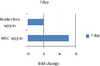 |
Folds Change During Expansion Under Different Conditions (w/cyto, with cytokine)
|
In this protocol, all CD3- cells such as monocytes were entered to the co-culture. These cells were actually contributed to the NK cells proliferation by cytokine secretion. This finding was consistent with the observation of Gada et al. (18), who reported that highly pure CD56+ cells from CB-MNCs had a poor expansion (only 4.5 fold) compared to CB-MNCs that were CD3-depleted only (14.5-fold).
In summary, our study showed that MSCs feeder incorporation with NK cells lineage specific cytokines could enhance fold expansion making it a suitable method to gain competent NK cells for immunotherapy.
Footnotes
References
- 1. Rosenberg SA, Yannelli JR, Yang JC, Topalian SL, Schwartzentruber DJ, Weber JS, et al. Treatment of patients with metastatic melanoma with autologous tumor-infiltrating lymphocytes and interleukin 2. J Natl Cancer Inst. 1994;86(15):1159-66. [PubMed]
- 2. Burnet FM. The Concept of Immunological Surveillance. Progress Exp Tumor Res. 1970;13:1-27. [DOI]
- 3. Wiltrout RH. Regulation and antimetastatic functions of liver-associated natural killer cells. Immunol Rev. 2000;174:63-76. [PubMed]
- 4. Herberman R. Ortaldo.. Natural killer cells: their roles in defenses against disease. Science. 1981;214(4516):24-30. [DOI]
- 5. Herberman RB, Nunn ME, Holden HT, Lavrin DH. Natural cytotoxic reactivity of mouse lymphoid cells against syngeneic and allogeneic tumors. II. Characterization of effector cells. Int J Cancer. 1975;16(2):230-9. [PubMed]
- 6. Cho D, Shook DR, Shimasaki N, Chang YH, Fujisaki H, Campana D. Cytotoxicity of activated natural killer cells against pediatric solid tumors. Clin Cancer Res. 2010;16(15):3901-9. [PubMed]
- 7. Geller MA, Cooley S, Judson PL, Ghebre R, Carson LF, Argenta PA, et al. A phase II study of allogeneic natural killer cell therapy to treat patients with recurrent ovarian and breast cancer. Cytotherapy. 2011;13(1):98-107. [PubMed]
- 8. Raulet DH, Held W. Natural killer cell receptors: The offs and ons of NK cell recognition. Cell. 1995;82(5):697-700. [DOI]
- 9. Karre K. Express yourself or die: peptides, MHC molecules, and NK cells. Science. 1995;267(5200):978-9. [PubMed]
- 10. Moretta A, Vitale M, Bottino C, Orengo AM, Morelli L, Augugliaro R, et al. P58 molecules as putative receptors for major histocompatibility complex (MHC) class I molecules in human natural killer (NK) cells. Anti-p58 antibodies reconstitute lysis of MHC class I-protected cells in NK clones displaying different specificities. J Exp Med. 1993;178(2):597-604. [PubMed]
- 11. McGuckin C, Jurga M, Ali H, Strbad M, Forraz N. Culture of embryonic-like stem cells from human umbilical cord blood and onward differentiation to neural cells in vitro. Nat Protoc. 2008;3(6):1046-55. [PubMed]
- 12. Pipes BL, Tsang T, Peng SX, Fiederlein R, Graham M, Harris DT. Telomere length changes after umbilical cord blood transplant. Transfusion. 2006;46(6):1038-43. [PubMed]
- 13. Olson JA, Leveson-Gower DB, Gill S, Baker J, Beilhack A, Negrin RS. NK cells mediate reduction of GVHD by inhibiting activated, alloreactive T cells while retaining GVT effects. Blood. 2010;115(21):4293-301. [PubMed]
- 14. Yu H, Fehniger TA, Fuchshuber P, Thiel KS, Vivier E, Carson WE, et al. Flt3 ligand promotes the generation of a distinct CD34(+) human natural killer cell progenitor that responds to interleukin-15. Blood. 1998;92(10):3647-57. [PubMed]
- 15. Shah AJ, Smogorzewska EM, Hannum C, Crooks GM. Flt3 ligand induces proliferation of quiescent human bone marrow CD34+CD38- cells and maintains progenitor cells in vitro. Blood. 1996;87(9):3563-70. [PubMed]
- 16. Fujisaki H, Kakuda H, Shimasaki N, Imai C, Ma J, Lockey T, et al. Expansion of highly cytotoxic human natural killer cells for cancer cell therapy. Cancer Res. 2009;69(9):4010-7. [PubMed]
- 17. Castro-Manrreza ME, Montesinos JJ. Immunoregulation by mesenchymal stem cells: biological aspects and clinical applications. J Immunol Res. 2015;2015:394917. [PubMed]
- 18. Gada P, Gleason M, McCullar V, McGlave PB, Miller JS, editor(s). Optimal NK Cell Expansion Depends on Accessory Cells, Synergy between Physiologic Concentrations of IL-2 and IL-15, and Umbilical Cord Blood (UCB) NK Cell Precursors Expand Better Than Adult NK Cells. ASH Annual Meeting Abstracts; 2006; 3642 p.